Understanding Gingival Pigmentation:
The color of the gums, or gingiva, varies among individuals and is influenced by surface pigmentation. It can range from light to dark brown or black, reflecting differences in skin tone, texture, and color across races and regions. Gingival color is primarily determined by factors such as blood supply, the thickness of the surface layer (epithelium), degree of keratinization, and various pigments present within the gingiva, including Melanin, Carotene, reduced haemoglobin, and oxy-haemoglobin.
Gingival pigmentation manifests as a diffused deep purplish discoloration or irregular brown to black patches, striae, or strands. This pigmentation is the result of melanin granules produced by melanoblast cells. Melanin, a brown pigment derived from non-haemoglobin sources, is primarily found in the basal and supra-basal cell layers of the epithelium.
Interestingly, melanin pigmentation can appear in oral tissues as early as 3 hours after birth and can sometimes be the sole indicator of pigmentation on the body. It’s widely accepted that pigmented areas form when melanin granules produced by melanocytes are transferred to keratinocytes, a relationship referred to as the ‘epidermal-melanin unit.’
Laser Gum Depigmentation:
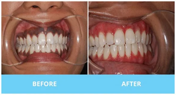
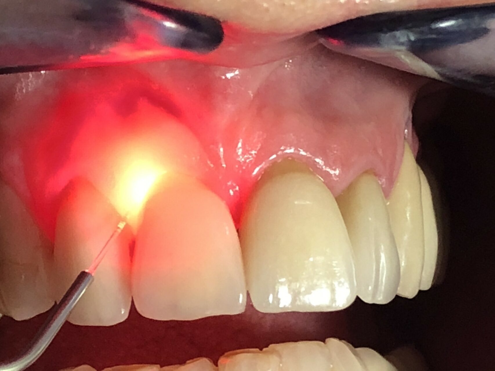
Laser gum depigmentation involves the use of a dental laser to remove or reduce the melanin pigmentation from the gums. This procedure offers several advantages, including precise control, minimal discomfort, and faster healing compared to manual methods.
Manual Gum Depigmentation:
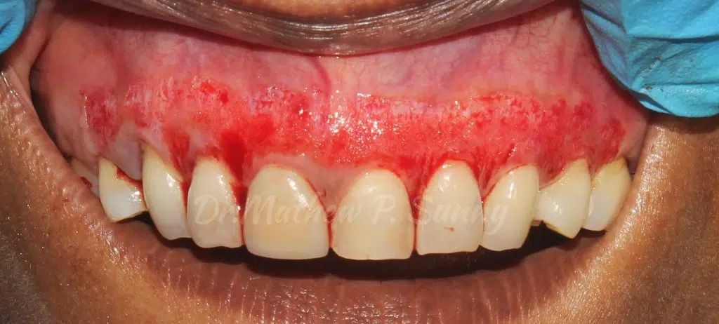
Manual gum depigmentation, also known as surgical gum depigmentation, involves using a scalpel or abrasive instruments to remove the pigmented gum tissue. While effective, this method may result in more post-operative discomfort and a longer healing period compared to laser gum depigmentation.


